Lewiston Formerly Institute of Physical Therapy
Lewiston, ID 83501 Get Directions
Phone
(208) 746-1418Fax
(208) 746-4123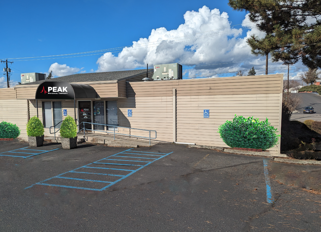
Location Info
This Location is Now Hiring
Apply Now!
Clinic Hours
- Monday
- 8:00 AM - 5:00 PM
- Tuesday
- 8:00 AM - 5:00 PM
- Wednesday
- 8:00 AM - 5:00 PM
- Thursday
- 8:00 AM - 5:00 PM
- Friday
- 8:00 AM - 5:00 PM
- Saturday
- Closed
- Sunday
- Closed
Meet the Team
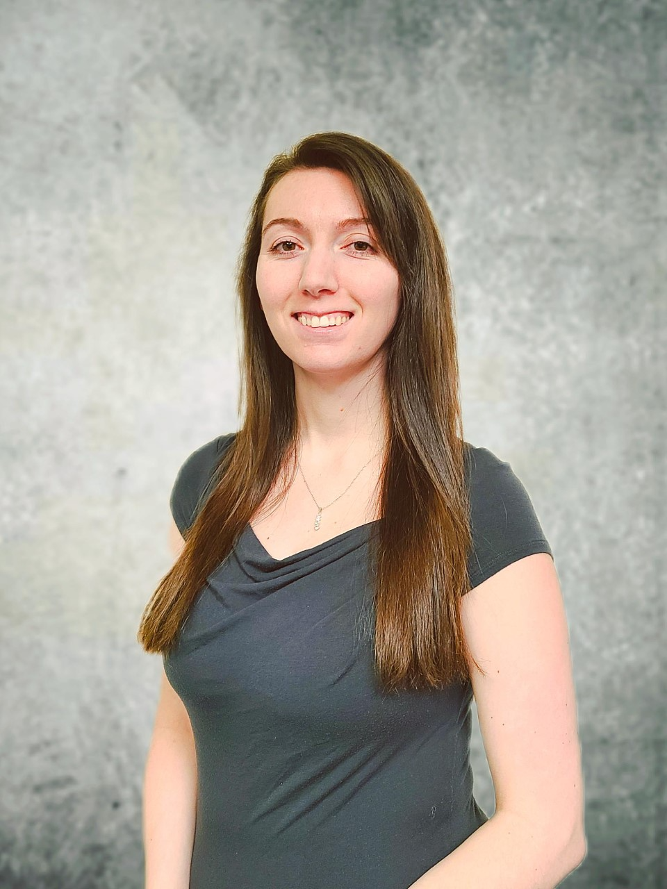
Levi Frasier PT, DPT
Clinic Director, Physical Therapist
Levi Frasier PT, DPT
Clinic Director, Physical TherapistLevi joined the Institute of Physical Therapy in 2008. He is a native of the Lewis-Clark Valley. He completed his Bachelor of Science in Physical Education at the University of Idaho and went on to receive his Doctorate of Physical Therapy from the University of Puget Sound. Through continuing education, he strives to provide the most recent treatment approaches in physical therapy tailored to each individual’s specific needs and goals.
Levi is also a lab instructor for the LCSC Physical Therapist Assistant (PTA) Program through the Idaho Consortium for PTA education. He thoroughly enjoys helping to educate the physical therapist assistant students.
In addition, Levi also enjoys many outdoor activities with his wife and two boys—camping, boating, water and snow skiing, and golfing.
Jaymes Sajczuk PT, DPT
Physical Therapist
Jaymes Sajczuk PT, DPT
Physical TherapistJaymes Sajczuk PT, DPT is a graduate of the Doctor of Physical Therapy program at Creighton University in 2020. Originally from Arvada, Colorado he met his wife at Colorado State University in Fort Collins, Colorado. They moved to Lewiston, Idaho from Colorado for the balance of the atmosphere of the town as well as the opportunities to be outdoors while escaping the big cities of Denver and Colorado Springs. They both love to be outdoors with their son and dog and are very active in their local church. Since moving to Lewiston, Jaymes has begun hunting and fishing, and is very passionate about the outdoors. Jaymes loves treating orthopedic cases but treating individuals with limb loss was what got him originally into physical therapy.
Shelby Holland PTA
Physical Therapy Assistant
Shelby Holland PTA
Physical Therapy AssistantShelby graduated from North Idaho College with a PTA degree in May 2022. Shelby and her husband have a beautiful baby boy and moved to Lewiston, Idaho in April 2020. She loves being outdoors, enjoys nature with her family and going on new adventures. Shelby loves anything Disney and dreams of taking her son to Disneyland to enjoy the happiest place on Earth! She loves working with all patients but has a special passion for pregnant and postpartum women, and helping them return to doing what they love after giving birth to a miracle.
Peak Physical Therapy in Nez Perce County
When searching for top-notch physical therapy clinics near St. Joseph Regional Medical Center in Lewiston, ID, look to Peak Physical Therapy (formerly known as the Institute of Physical Therapy). Conveniently located in Nez Perce County in Lewiston, we are situated just slightly east of Rte.129 and South of Rte.12. We are an exceptional team of physical therapists near Lewiston-Ned Perce County Regional Airport who offer cutting-edge treatments for a wide variety of conditions. We are the perfect choice for anyone seeking physical therapy near Potlatch Credit Union. Our professional and dedicated team takes pride in delivering effective and advanced patient care in a comfortable and welcoming environment. Call or visit us online to schedule your initial screening visit. Let Peak PT help you “Reclaim Your Life” with a personalized treatment plan to alleviate your discomfort with long-term sustainable results.
Specialties & Treatments at Our Clinic
Count on the dedicated therapists at Peak Physical Therapy in Lewiston to offer exceptional patient care tailored to your needs. Whether you’re dealing with chronic pain from a traumatic injury or recovering from surgery, our highly trained physical therapists are here to support you throughout every stage of your rehabilitation. Our professional team will assist you in achieving a pain-free and optimal physical health and mobility level. Our Lewiston Peak PT clinic provides diverse treatments, specializing in Back Pain Rehabilitation, Vestibular Therapy, TMJ Therapy, Sports Rehabilitation, and Orthopedic Therapy. We also offer techniques like Manual Therapy that focus on managing musculoskeletal conditions. Your first steps toward ‘Maximal Functional Results’ start at Peak Physical Therapy in Lewiston—partner with us for a successful recovery with the long-term results you deserve.
Meet Our Top Physical Therapists in Lewiston
At Peak Physical Therapy in Lewiston, ID, our team of degreed and certified physical therapists is dedicated to staying at the forefront of physical therapy research, techniques, and equipment. We believe in building meaningful relationships with our patients, understanding that effective therapy and compassionate care are crucial to healing. By addressing the root cause of your concerns and providing ongoing support, we strive to enhance your quality of life and reduce healthcare costs. We aim to help you achieve a healthier, pain-free, and active lifestyle. Schedule your first appointment with us today, and let our team assist you every step of the way, from appointment scheduling to insurance claims and payment plans. Your journey to recovery starts here at Peak Physical Therapy.
People in Idaho and Washington Trust Peak PT
Hear from patients that reclaimed their lives.